Liver Disease Blood Test
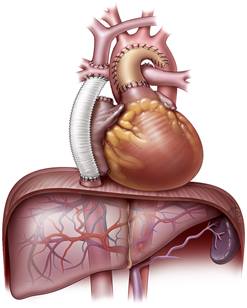
There has been growing awareness of the development of liver disease in Fontan patients. This is thought to be the result of a number of factors, including multiple surgeries, medications, overfilling of the veins with blood, and volume overload on the liver due to the lack of a ventricle to pump blood to the lung arteries. Blood tests and imaging studies
are used for ongoing monitoring because they are low-to noninvasive. The clinic uses a combination of blood and imaging tests to identify patients who should undergo a liver biopsy, which is an invasive test.
- Liver Function/Enzyme Tests
These blood tests help to determine liver health by measuring the levels of certain proteins and enzymes in the blood.
- FibroSure
This noninvasive assessment of liver fibrosis analyzes 10 biochemicals in combination with age, gender, height, and weight.
- Complete Blood Count (CBC)
CBC is one of the most commonly ordered blood tests because liver damage can affect the blood count.
- Viral Hepatitis Test
Viral hepatitis can be a cause of liver dysfunction.
- Alpha-Fetoprotein (AFP) Blood Test
AFP is a protein normally produced by the liver and is used for screening of liver disease and protein-losing diseases of the intestines.
Liver Biopsy
Liver biopsy remains the gold standard for assessment of liver disease. A liver biopsy is a procedure using a needle to remove a small piece of the liver so that it can be examined under a microscope for signs of damage or disease. The test is conducted in a hospital as an outpatient procedure. Before the test is administered, patients are given medicines for sedation and pain. It is important for patients to remain as still as possible during the biopsy, which is commonly done through the abdominal wall with patients lying on their backs. After cleaning and numbing the area, a biopsy needle is inserted. Ultrasound is typically
used to guide the needle or mark an optimal location. After the biopsy is obtained, the needle is removed quickly and a bandage is placed over the insertion site. The biopsy can also be done by inserting a needle into the jugular vein during cardiac catheterization. If the procedure is performed this way, X-rays will be used to guide the needle to the vein. A special needle and catheter is used to take the biopsy sample. Patients will need someone to drive them home.

Liver MRI & CT Scans
MRI scans provide detailed images of soft tissues in the body by using radio waves and strong magnets instead of X-rays. When MRI is used to look at the liver, several sets of images may be taken. After the first set is done, a contrast material called gadolinium is injected into a vein
in order to help show details more clearly. New sets of images are taken over the next several minutes as the gadolinium moves through the liver and other parts of the body. This is known as dynamic contrast-enhanced MRI, which is beneficial for viewing liver abnormalities like fibrosis and cirrhosis, and can help physicians discern between benign and malignant tumors. The scans can also be used to look at blood vessels in and around the liver.
For some patients, including those with metal implants such as a pacemaker or an implantable cardioverter-defibrillator (ICD), MRI is not feasible. These patients will have their livers evaluated with a CT scan. A CT scan is an X-ray test that produces detailed cross-sectional images
of the body. Similar to MRI, CT scans are helpful in looking at liver abnormalities like fibrosis, cirrhosis, and both non-malignant and malignant tumors, as well as nearby blood vessels.